
LOGIQ™
FAQs
Get all the information you need
LOGIQ™
FAQs
Get all the information you need
What are the benefits of ultrasound in Liver disease management?
Ultrasound is a valuable tool in the diagnosis and management of liver diseases due to its non-invasiveness, cost-effectiveness, versatility, real-time imaging capabilities, and minimal preparation requirements:
- Cost-effectiveness: Ultrasound is a relatively inexpensive diagnostic tool compared to other imaging techniques such as CT or MRI scans, making it more accessible for patients and healthcare systems.
- Versatility: Ultrasound can be used to diagnose a wide range of liver diseases, including liver cancer, cirrhosis, and fatty liver disease, among others.
- Real-time imaging: Ultrasound provides real-time imaging of the liver, allowing healthcare providers to monitor changes in the liver over time and adjust treatment plans accordingly.
- Minimal preparation: Unlike some other imaging techniques, ultrasound does not require significant preparation such as fasting or contrast dyes, making it a more convenient option for patients.
How can ultrasound accompany the liver care pathway?
Ultrasound can accompany the full liver care pathway by assisting in diagnosis, treatment planning, monitoring, screening, and biopsy guidance:
- Diagnosis: Ultrasound can be used to diagnose various liver diseases such as fatty liver disease, cirrhosis, and liver cancer. Early detection of these diseases is critical in providing appropriate treatment and improving patient outcomes.
- Treatment planning: Ultrasound can assist in treatment planning by providing real-time imaging of the liver and surrounding organs. This allows healthcare providers to assess the extent of liver damage and plan appropriate interventions, such as medication or surgery.
- Monitoring: Ultrasound can be used to monitor changes in the liver over time, allowing healthcare providers to track disease progression and adjust treatment plans accordingly.
- Screening: Ultrasound can also be used as a screening tool for individuals at risk of liver disease, such as those with a history of heavy alcohol consumption or viral hepatitis. Regular ultrasound screenings can help detect liver disease in its early stages, when treatment is most effective.
- Biopsy guidance: Ultrasound can guide liver biopsies, which are used to obtain liver tissue samples for diagnosis and treatment planning. Ultrasound guidance improves the accuracy and safety of liver biopsies.
How can Ultrasound-Guided Attenuation Parameter (UGAP) support the quantification of liver steatosis?
UGAP measures the attenuation or weakening of ultrasound signals as they pass through the liver, and this attenuation is correlated with the amount of fat in the liver, allowing for the quantification of liver steatosis with accuracy similar to histology and MRI-PDFF.
What are the advantages of contrast-enhanced ultrasound (CEUS) in liver imaging?
Contrast-enhanced ultrasound has several advantages in liver imaging. First, it provides real-time imaging of liver vasculature, allowing for accurate diagnosis of liver lesions and tumors. Second, it is a non-invasive procedure that does not involve ionizing radiation or contrast agents, making it a safer alternative to other imaging techniques. Finally, CEUS is highly sensitive to changes in blood flow and perfusion, making it a valuable tool in the diagnosis and treatment of liver diseases such as cirrhosis and liver cancer.
What are the benefits of ultrasound in Musculoskeletal disease management?
Ultrasound is a valuable tool in the management of musculoskeletal diseases. It is safe, accurate, cost-effective, and provides real-time imaging, making it a preferred option for many patients and healthcare providers:
- Non-invasive: Ultrasound is a non-invasive diagnostic tool, which means it doesn't require any surgical incisions or injections. This makes it a safer and less painful option for patients.
- Real-time imaging: Unlike X-rays or MRI scans, which provide static images, ultrasound provides real-time images of the affected area. This allows physicians to observe the movement and function of muscles, tendons, and other soft tissues in real-time.
- Accuracy: Ultrasound has been shown to be highly accurate in diagnosing musculoskeletal conditions such as rotator cuff tears, carpal tunnel syndrome, and plantar fasciitis. It can also help physicians identify other conditions that may be causing pain or discomfort.
- Cost-effective: Compared to other imaging techniques, such as MRI scans, ultrasound is generally more cost-effective. This makes it a good option for patients who may not be able to afford more expensive imaging tests.
- Guided injections: Ultrasound can also be used to guide injections of medication directly into the affected area. This allows physicians to precisely target the source of pain or inflammation, resulting in better outcomes for patients.
In which musculoskeletal applications and how does ultrasound help in the diagnosis and management of musculoskeletal disorders?
Ultrasound is a valuable tool in the diagnosis and management of various musculoskeletal disorders. It can be used to visualize soft tissue structures such as muscles, tendons, and ligaments, as well as bones and joints. Ultrasound can help identify and diagnose conditions such as tendonitis, carpal tunnel syndrome, and rotator cuff tears. Additionally, ultrasound-guided interventions such as injections and aspirations can be performed to manage pain and inflammation. Ultrasound is a safe, non-invasive, and cost-effective imaging modality that can be used in various settings, including clinics and sports medicine facilities.
How can advanced solutions as Elastography and Flow Quantification support musculoskeletal ultrasound assessments?
Advanced solutions such as Elastography and Flow Quantification can enhance musculoskeletal ultrasound assessments in several ways. Elastography can help evaluate tissue stiffness and elasticity, which can aid in the diagnosis and monitoring of various musculoskeletal conditions such as tendon injuries, muscle tears, and fibrotic changes. It can also provide information about changes in tissue over time, which can help in treatment planning and management. Flow quantification, on the other hand, can provide information about blood flow in the examined area, which can be useful in the diagnosis and management of vascular and musculoskeletal conditions. By incorporating these advanced solutions into musculoskeletal ultrasound assessments, healthcare providers can obtain additional information that can improve the accuracy and reliability of their diagnoses and treatment plans.
How can ultrasound accompany the musculoskeletal care pathway?
- Ultrasound can be used as a non-invasive diagnostic tool to identify and diagnose musculoskeletal conditions such as tendonitis, muscle tears, and joint disorders.
- Ultrasound-guided interventions such as injections and aspirations can be performed to manage pain and inflammation.
- Ultrasound can be used to monitor the progression of musculoskeletal disorders and the effectiveness of treatments over time.
- Finally, ultrasound can also be used for pre-operative planning and post-operative follow-up in orthopedic surgeries.
By incorporating ultrasound into the musculoskeletal care pathway, healthcare providers can improve the accuracy of their diagnoses, enhance treatment planning and management, and improve patient outcomes.
What are the benefits of ultrasound in breast disease management?
Ultrasound is a valuable tool in the management of breast disease. It is safe, accurate, cost-effective, and provides real-time imaging, making it a preferred option for many patients and healthcare providers:
- Non-invasive: Ultrasound is a non-invasive imaging technique, which means it doesn't require any surgical incisions or injections. This makes it a safer and less painful option for patients.
- High accuracy: Ultrasound has been shown to be highly accurate in identifying and characterizing breast lesions. It can distinguish between benign and malignant tumors and can also be used to guide biopsies.
- Real-time imaging: Ultrasound provides real-time images of the breast tissue, which allows physicians to observe the movement and function of tissues in real-time. This can be particularly useful for evaluating changes over time or for monitoring treatment responses.
- Cost-effective: Compared to other imaging techniques, such as MRI scans, ultrasound is generally more cost-effective. This makes it a good option for patients who may not be able to afford more expensive imaging tests.
- Safe for pregnant women: Ultrasound is safe for pregnant women, which makes it a preferred imaging option for evaluating breast conditions in pregnant or breastfeeding women.
How can ABUS support the detection of small invasive cancers in dense breast when used as adjunct to mammography?
Automated breast ultrasound (ABUS) can support the detection of small invasive cancers in dense breast tissue when used as an adjunct to mammography. Dense breast tissue can make it more difficult to detect small cancers with mammography alone. ABUS can provide additional images of the breast tissue, which can help identify small cancers that may be missed on mammography. Studies have shown that ABUS can increase cancer detection rates in women with dense breast tissue, particularly for small invasive cancers. By using ABUS as an adjunct to mammography, healthcare providers can improve the accuracy of their breast cancer screening and potentially detect cancers at an earlier stage, leading to better patient outcomes.
How can ABUS and ultrasound help deliver a personalized breast care to the patients, across the care pathway?
By incorporating ABUS and ultrasound into breast care pathways, healthcare providers can personalize breast care for each patient, improving accuracy, reducing unnecessary procedures, and ultimately improving patient outcomes:
- Screening: ABUS and ultrasound can be used as adjuncts to mammography in breast cancer screening, particularly in women with dense breast tissue. By providing additional imaging information, healthcare providers can personalize screening protocols for each patient based on their individual risk factors and breast tissue characteristics.
- Diagnosis: Ultrasound can be used to diagnose breast conditions, such as cysts, lumps, and fibroadenomas, which may not be visible on mammography. ABUS can also be used to improve diagnostic accuracy.
- Treatment planning: Ultrasound can be used to guide biopsies, aspiration, and other interventional procedures, which can help personalize treatment planning for each patient based on their individual needs.
- Follow-up: Ultrasound can be used to monitor patients following breast cancer treatment, helping to detect any signs of recurrence or other complications. This can help personalize follow-up care plans for each patient based on their individual history and risk factors.
Discover our clinical tutorials by care area
Go to clinical education

Interested in requesting a quotation?
Configure my LOGIQ
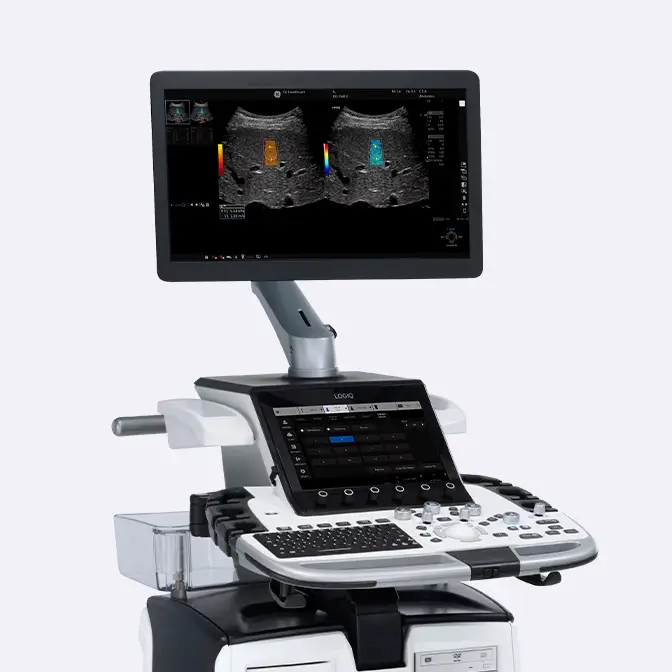
LOGIQ E10 Series
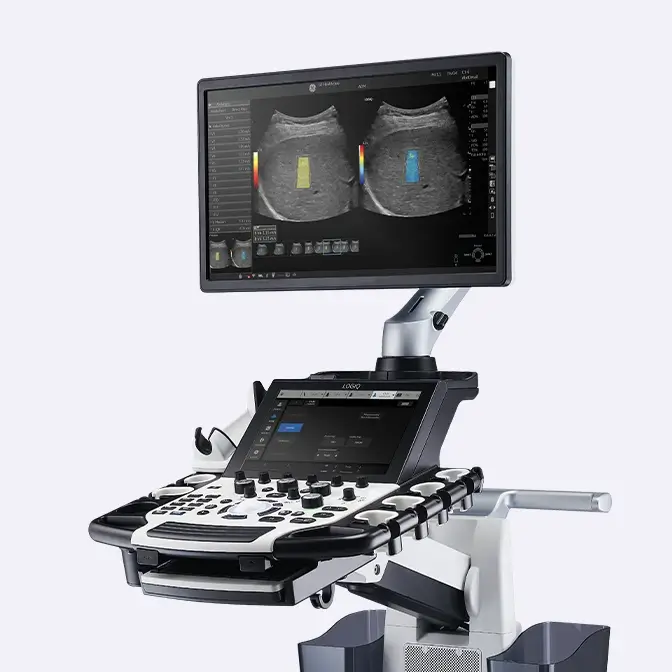
LOGIQ Fortis
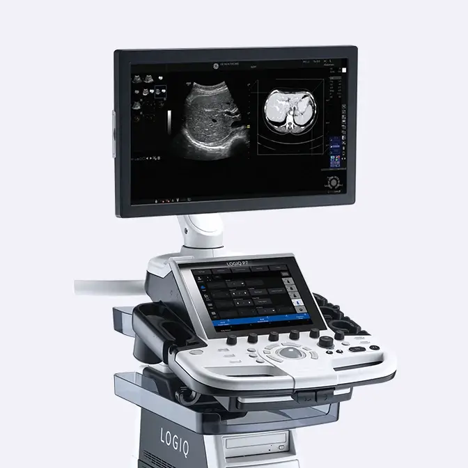
LOGIQ P Series
Have a question?
We would love to hear from you.
Contact us 


