
Managing breast care with ultrasound
Personalized breast care
with ultrasound
Managing liver care with ultrasound
Personalized breast care
with ultrasound
Screening
The power of early cancer detection
Supplemental screening with Invenia ABUS transforms breast care from reactive to proactive. Clinical studies have demonstrated that ABUS can help increase the detection of small invasive cancers in dense breasts by 57% when used as adjunct to mammography1. Detection cancers at early stage has important prognostic implications.
1. Wilczek, et.al. Adding 3D Automated Breast Ultrasound to mammography screening in women with heterogeneously and extremely dense breasts. European Journal of Radiology 85 (2016) 1554–1563
1. Wilczek, et.al. Adding 3D Automated Breast Ultrasound to mammography screening in women with heterogeneously and extremely dense breasts. European Journal of Radiology 85 (2016) 1554–1563

Invenia ABUS is the first FDA approved screening Ultrasound device.
Automated Breast Ultrasound improve early cancer detection with an automated, user-independent and reproducible screening solution for dense breasts and high-risk screening programs.
Learn more about the ABUS technology Learn more about Invenia ABUS key considerations Learn how to use 3D ABUS to achieve improved breast care outcomes Invenia ABUS 2.0 - Standard Acquisition Testimonial - Personalized breast imaging Diagnosis/Staging
Characterize lesion, access spread
Perform an accurate diagnosis and staging of a lesion is key to implement the best treatment plan. The latest ultrasound innovations and AI-based solutions help elevate diagnostic with higher accuracy and increased clinical confidence.

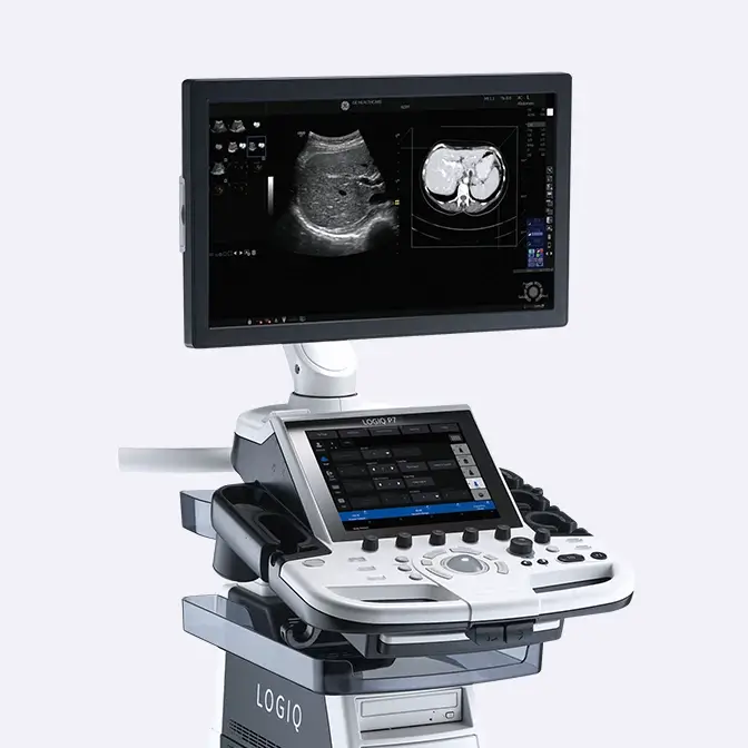
Diagnosis B-MODE
B-Mode Image quality is a key feature in Ultrasound driving confidence, workflow and serving as enabler for expert tools. Innovative system architectures, equipped with the latest imaging processing techniques (as the cSound™ Imageformer), the e-Series Matrix probe technology and imaging display allow to optimize the signal along the imaging process from probe to pixel. In Breast, this results in an image with exquisite spatial and contrast resolution, focused from near to far field, enabling a clear depiction and analysis of the smallest tissue abnormalities.
Learn more about the advantages of the cSound Imaging Technology 
Flow Modes
A comprehensive package of Flow Modes, including the latest Micro-Vascular Imaging with Radiantflow™ and B-Flow Imaging, allow to see and assess vascularity at an earlier stage of the disease progression.
Learn which is the LOGIQ Vascular solution fitting your needs 
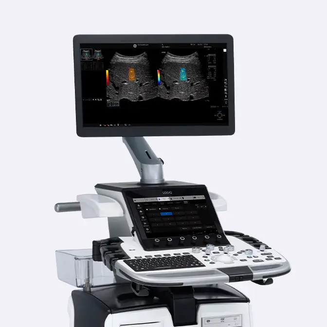
2D Shear Wave Elastography (2D SWE)
There is a need of non-invasive techniques to support you in lesions classification. 2D SWE is a technique helping to support decisions through the assessment of tissue stiffness. Easy measurement of propagation speed of shear waves help get more information on the masses structure, increasing the confidence in the characterization of lesions being benign or malignant. 2D SWE is available on multiple probes and can run both on live examination and raw data.
Learn more about the LOGIQ 2D SWE technique 
Decision Support with Koios DS
Breast Assistant, powered by Koios DS is an AI-based decision support tool. The solution automatically provides an AI-based quantitative risk assessment that aligns to a BI-RADS ATLAS® category helping improve consistency across users and reduce unnecessary biopsies.
Learn more about LOGIQ Breast Assistant Read the details of Clinical Study supporting Koios DS FDA registration Watch how Breast Assistant powered by Koios DS works 
See more with multi-modality imaging
The easy comparison of ultrasound images with acquired images or dataset from other modalities is important to increase the level of information and help guide the user to the exact region of interest.
Learn more about LOGIQ Breast Assistant 
Automated Breast Ultrasound
ABUS provides standardized, repeatable observations and double reading possibilities within multiple volumes leading into great advantages for multifocal and multi-centric cancer disease management.
Learn more about the ABUS technology Learn more about Invenia ABUS key considerations Learn how to use 3D ABUS to achieve improved breast care outcomes Invenia ABUS 2.0 - Standard Acquisition Testimonial - Personalized breast imaging B-Mode
Diagnosis B-MODE
B-Mode Image quality is a key feature in Ultrasound driving confidence, workflow and serving as enabler for expert tools. Innovative system architectures, equipped with the latest imaging processing techniques (as the cSound™ Imageformer), the e-Series Matrix probe technology and imaging display allow to optimize the signal along the imaging process from probe to pixel. In Breast, this results in an image with exquisite spatial and contrast resolution, focused from near to far field, enabling a clear depiction and analysis of the smallest tissue abnormalities.
Learn more about the advantages of the cSound Imaging Technology Flow Modes
Flow Modes
A comprehensive package of Flow Modes, including the latest Micro-Vascular Imaging with Radiantflow™ and B-Flow Imaging, allow to see and assess vascularity at an earlier stage of the disease progression.
Learn which is the LOGIQ Vascular solution fitting your needs Lesion assessment with 2DSWE
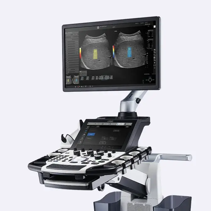
2D Shear Wave Elastography (2D SWE)
There is a need of non-invasive techniques to support you in lesions classification. 2D SWE is a technique helping to support decisions through the assessment of tissue stiffness. Easy measurement of propagation speed of shear waves help get more information on the masses structure, increasing the confidence in the characterization of lesions being benign or malignant. 2D SWE is available on multiple probes and can run both on live examination and raw data.
Learn more about the LOGIQ 2D SWE technique Decision Support with Koios DS™
Decision Support with Koios DS
Breast Assistant, powered by Koios DS is an AI-based decision support tool. The solution automatically provides an AI-based quantitative risk assessment that aligns to a BI-RADS ATLAS® category helping improve consistency across users and reduce unnecessary biopsies.
Learn more about LOGIQ Breast Assistant Read the details of Clinical Study supporting Koios DS FDA registration Watch how Breast Assistant powered by Koios DS works Multi-modality imaging
See more with multi-modality imaging
The easy comparison of ultrasound images with acquired images or dataset from other modalities is important to increase the level of information and help guide the user to the exact region of interest.
Learn more about LOGIQ Breast Assistant Automated breast ultrasound
Automated Breast Ultrasound
ABUS provides standardized, repeatable observations and double reading possibilities within multiple volumes leading into great advantages for multifocal and multi-centric cancer disease management.
Learn more about the ABUS technology Learn more about Invenia ABUS key considerations Learn how to use 3D ABUS to achieve improved breast care outcomes Invenia ABUS 2.0 - Standard Acquisition Testimonial - Personalized breast imaging Treatment
Planning, monitoring
To implement the best personalized treatment for each of your patients is important to success. You can find advanced solutions to support the most effective and less invasive treatment for every individual woman to perform an accurate planning, guidance and monitoring.

B-Steer+
To increase your confidence in needle guidance, B-Steer+ enhances needle visualisation in real time, improving speed and confidence in needle guidance procedures. Download Icon
Learn more about LOGIQ Breast Assistant 
2D Shear Wave Elastography (2D SWE)
Elastography may increase your confidence in treatment plan. It can help determine the actual size of the breast lesion and the extent of tissue infiltration, driving the best approach in surgical procedures.
Learn more about the LOGIQ 2D SWE technique 
Volume Navigation (V Nav) with Fusion Imaging
How many information can be collected combining different imaging modalities? Fusion Imaging US/MRI facilitates a second look examination and allows guidance of biopsies for lesions seen on MRI but not on US. This allows to have a better patient experience, avoiding time consuming and expensive MRI guided biopsies.
Clinical How To presentation by Dr A Fausto at EUSOBI 2019 Clinical presentation by Dr A. Fausto at ECR 2019 Clinical case explanation by Mrs Robina Parvez 
Automated Breast Ultrasound
ABUS provides a full view of the breast, depicting the extent of lesion, multiplicity and potential stromal involvement helping in a better surgical approach. It can be also important in the margin control of surgical specimen, to improve accuracy in breast conserving surgery.
Learn more about the ABUS technology Learn more about Invenia ABUS key considerations Learn how to use 3D ABUS to achieve improved breast care outcomes Invenia ABUS 2.0 - Standard Acquisition Testimonial - Personalized breast imaging B-Steer+
B-Steer+
To increase your confidence in needle guidance, B-Steer+ enhances needle visualisation in real time, improving speed and confidence in needle guidance procedures. Download Icon
Learn more about LOGIQ Breast Assistant 2D Shear Wave Elastography
2D Shear Wave Elastography (2D SWE)
Elastography may increase your confidence in treatment plan. It can help determine the actual size of the breast lesion and the extent of tissue infiltration, driving the best approach in surgical procedures.
Learn more about the LOGIQ 2D SWE technique US/MRI Fusion
Volume Navigation (V Nav) with Fusion Imaging
How many information can be collected combining different imaging modalities? Fusion Imaging US/MRI facilitates a second look examination and allows guidance of biopsies for lesions seen on MRI but not on US. This allows to have a better patient experience, avoiding time consuming and expensive MRI guided biopsies.
Clinical How To presentation by Dr A Fausto at EUSOBI 2019 Clinical presentation by Dr A. Fausto at ECR 2019 Clinical case explanation by Mrs Robina Parvez Automated Breast Ultrasound
Automated Breast Ultrasound
ABUS provides a full view of the breast, depicting the extent of lesion, multiplicity and potential stromal involvement helping in a better surgical approach. It can be also important in the margin control of surgical specimen, to improve accuracy in breast conserving surgery.
Learn more about the ABUS technology Learn more about Invenia ABUS key considerations Learn how to use 3D ABUS to achieve improved breast care outcomes Invenia ABUS 2.0 - Standard Acquisition Testimonial - Personalized breast imaging Follow up
Complications & recurrences
It is key to support breast care patients in the follow up of their interventions or drug therapies, to avoid any potential complication and minimize their risk of recurrences. It is also important to monitor any potential mass that is kept under observation. This would help to ensure a better patient experience with a non-invasive, patient-friendly and low cost technology providing advanced auto prior compare features.

Treatment response with Contrast-Enhanced Ultrasound (CEUS)
CEUS could help in the characterization of lesions based on the different contrast uptake times and in the assessment of NCA treatment response.
Clinical Web Series presentation - Dr E. Divjak 
Automated Breast Ultrasound (ABUS)
A non-invasive and precise follow-up method: The 3D and coronal plane access, combined with the Auto Prior function, allows an accurate diagnosis and a standardized comparison with previous findings
Learn more about the ABUS technology Learn more about Invenia ABUS key considerations Learn how to use 3D ABUS to achieve improved breast care outcomes Invenia ABUS 2.0 - Standard Acquisition Testimonial - Personalized breast imaging Contrast-Enhanced Ultrasound
Treatment response with Contrast-Enhanced Ultrasound (CEUS)
CEUS could help in the characterization of lesions based on the different contrast uptake times and in the assessment of NCA treatment response.
Clinical Web Series presentation - Dr E. Divjak Automated Breast Ultrasound
Automated Breast Ultrasound (ABUS)
A non-invasive and precise follow-up method: The 3D and coronal plane access, combined with the Auto Prior function, allows an accurate diagnosis and a standardized comparison with previous findings
Learn more about the ABUS technology Learn more about Invenia ABUS key considerations Learn how to use 3D ABUS to achieve improved breast care outcomes Invenia ABUS 2.0 - Standard Acquisition Testimonial - Personalized breast imaging Back to breast care area site
Discover our clinical tutorials by care area
Go to clinical education

Interested in requesting a quotation?
Configure my LOGIQ
LOGIQ E10 Series
LOGIQ Fortis
LOGIQ P Series
Have a question?
We would love to hear from you.
Contact us 


