
Managing musculoskeletal care with ultrasound
See and quantify musculoskeletal
disorders in a single exam
Managing musculoskeletal care with ultrasound
See and quantify musculoskeletal disorders in a single exam
Diagnosis/Staging
Clinical challenges
Perform an accurate diagnosis and staging of a musculoskeletal condition is key to implement the best therapy and hopefully have a quick recovery of the patient.
Solutions
With the benefit of high-resolution B-Mode imaging, new Flow Modes being even more sensitive, and a portfolio of specialty probes, dynamic Ultrasound become the elected modality in the evaluation of ligaments, tendons, muscles, and nerves in the extremities. Advanced solutions as Elastography and Flow Quantification can also provide functional information and impact on the choice of the best treatment. The more focused the clinical question is, the more beneficial MSK US are in clinical practice.

See more with B-Mode
High B-Mode Image Quality is the key feature in MSK ultrasound driving diagnostic confidence, especially with tendon tears, peripheral neuropathies or ligament disruptions. The benefit of high-resolution imaging with an extended bandwidth is ideal for the study of a variety of musculoskeletal conditions. Advanced SRI and imaging processing techniques increase even further contrast and spatial resolution.
Learn more about the GE HealthCare LOGIQ™ musculoskeletal solutions Learn about the experience with high frequency imaging and Flow Modes from Dr G. Allen 
Early vascular evaluatios with Flow Modes
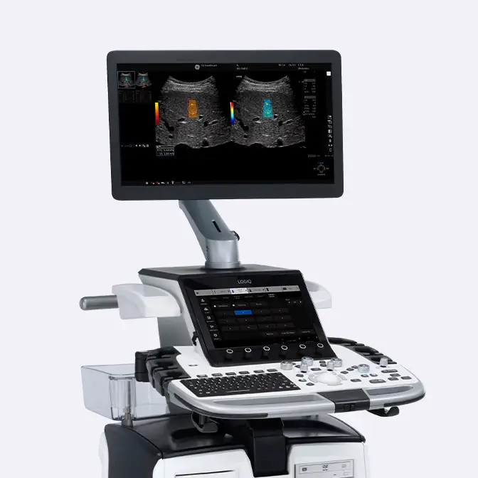
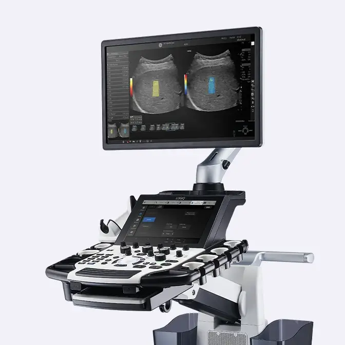
New Flow Modes, as MVI with Radiantflow™, are able to detect and display micro vessels and show their interconnection in a tissue. These techniques help in the study of tissue vascularity as in the assessment of potential inflammatory conditions (e.g. in rheumatology) potentially allowing an earlier staging of the disease process.
Learn more about the GE LOGIQ™ Vascular solutions Learn about the experience with High frequency Imaging and Flow Modes from Dr G. Allen 
2D SWE for tissue stiffness assessment
2D Shear Wave Elastography helps assess soft tissue stiffness in the study of tendons, ligaments and muscles injuries. It can also be used for the assessment of inflammatory disease, trauma extension in preparation of a recovery plan for athletes, or focal masses.
Learn more about the existing Elastography solutions 
A probe solution for every need
Latest innovations include high frequency probes, from large footprints to light hockey sticks, high performing low frequency, extended bandwidths and sensor embedded solutions for navigation. Delivering the needed information from neurology and rheumatology to soft tissue imaging.
Learn more about the last generation of GE LOGIQ probe solutions Learn about the experience with High frequency Imaging and Flow Modes from Dr G. Allen 
Inflammation staging with Flow quantification
2D Flow quantification delivers a semi-quantitative information of the vascular pattern in a selected ROI, helping stage the inflammatory disease key to setup a patient therapy.
Learn how to use Flow Quantification B-Mode
See more with B-Mode
High B-Mode Image Quality is the key feature in MSK ultrasound driving diagnostic confidence, especially with tendon tears, peripheral neuropathies or ligament disruptions. The benefit of high-resolution imaging with an extended bandwidth is ideal for the study of a variety of musculoskeletal conditions. Advanced SRI and imaging processing techniques increase even further contrast and spatial resolution.
Learn more about the GE HealthCare LOGIQ™ musculoskeletal solutions Learn about the experience with high frequency imaging and Flow Modes from Dr G. Allen Flow Modes
Early vascular evaluatios with Flow Modes
New Flow Modes, as MVI with Radiantflow™, are able to detect and display micro vessels and show their interconnection in a tissue. These techniques help in the study of tissue vascularity as in the assessment of potential inflammatory conditions (e.g. in rheumatology) potentially allowing an earlier staging of the disease process.
Learn more about the GE LOGIQ™ Vascular solutions Learn about the experience with High frequency Imaging and Flow Modes from Dr G. Allen 2D Shear Wave Elastography
2D SWE for tissue stiffness assessment
2D Shear Wave Elastography helps assess soft tissue stiffness in the study of tendons, ligaments and muscles injuries. It can also be used for the assessment of inflammatory disease, trauma extension in preparation of a recovery plan for athletes, or focal masses.
Learn more about the existing Elastography solutions A probe solution for every need
A probe solution for every need
Latest innovations include high frequency probes, from large footprints to light hockey sticks, high performing low frequency, extended bandwidths and sensor embedded solutions for navigation. Delivering the needed information from neurology and rheumatology to soft tissue imaging.
Learn more about the last generation of GE LOGIQ probe solutions Learn about the experience with High frequency Imaging and Flow Modes from Dr G. Allen Inflammation staging with Flow quantification
Inflammation staging with Flow quantification
2D Flow quantification delivers a semi-quantitative information of the vascular pattern in a selected ROI, helping stage the inflammatory disease key to setup a patient therapy.
Learn how to use Flow Quantification Treatment
Clinical challenges
Ultrasound are an ideal modality for image-guided MSK interventions, from planning to guidance. Techniques particularly pertinent to sports medicine include tendon fenestration/prolotherapy for the treatment of tendinopathy, injection of muscle tears, and perineural interventions, including hydro dissection.
Solutions
An advanced feature increasing the procedure confidence and success is B-Steer+, a technique enhancing the Needle visualization during the biopsy. Other invasive procedures include US image-guided biopsy of bone lesions, biopsy of MSK soft tissue masses and evaluation of MSK injury. Fusion imaging can be utilized in sacroiliac and facet joint injections, with additional confidence thanks to Needle Tip Tracking techniques. This could be particularly beneficial in young patients with inflammatory arthropathy, reducing the exposure to ionizing radiation.

Enhanced needle visualization with B-Steer+
In invasive procedures, B-Steer+ enhances needle visualization in real time, improving confidence during the procedure and saving time during biopsies and joint injections.
Learn more about the GE LOGIQ P Series Interventional solutions Learn more about the GE LOGIQ E10 Series Interventional solutions 
Precise guidance with Fusion Imaging
Volume Navigation (V Nav) with fusion Imaging, US/MRI or US/CT, can facilitate image-guided biopsy of bone lesions and soft tissue masses. Moreover, it is successfully utilized since years in sacroiliac and facet joint injections in combination with Needle Tip Tracking techniques.
Learn more about GE LOGIQ Interventional solutions Watch the experience from Dr Wilson in Oxford (ECR 2019 workshop) Increase biopsy confidence with Needle Tip Tracking
Needle Tip Tracking helps find the best needle path and follow the trajectory to the target. The technique helps Increase clinical confidence and accuracy in difficult procedures, where precision is key.
Learn more about GE LOGIQ Interventional solutions Watch the experience from Dr Wilson in Oxford (ECR 2019 workshop) 
Comprehensive information with Photo Assistant
Photo Assistant helps complete the examination with more visual information to be shared with referrals or peers. Acquire a photo of the relevant anatomy via an Android® device. The picture will be automatically transmitted to the ultrasound system and displayed in a side-by-side format with the live Ultrasound image, and saved to the patient exam data.
Watch how Photo Assistant works Read the experience using Photo Assistant from your peers B-Steer+
Enhanced needle visualization with B-Steer+
In invasive procedures, B-Steer+ enhances needle visualization in real time, improving confidence during the procedure and saving time during biopsies and joint injections.
Learn more about the GE LOGIQ P Series Interventional solutions Learn more about the GE LOGIQ E10 Series Interventional solutions Fusion Imaging
Precise guidance with Fusion Imaging
Volume Navigation (V Nav) with fusion Imaging, US/MRI or US/CT, can facilitate image-guided biopsy of bone lesions and soft tissue masses. Moreover, it is successfully utilized since years in sacroiliac and facet joint injections in combination with Needle Tip Tracking techniques.
Learn more about GE LOGIQ Interventional solutions Watch the experience from Dr Wilson in Oxford (ECR 2019 workshop) Needle Tip Tracking
Increase biopsy confidence with Needle Tip Tracking
Needle Tip Tracking helps find the best needle path and follow the trajectory to the target. The technique helps Increase clinical confidence and accuracy in difficult procedures, where precision is key.
Learn more about GE LOGIQ Interventional solutions Watch the experience from Dr Wilson in Oxford (ECR 2019 workshop) Photo Assistant
Comprehensive information with Photo Assistant
Photo Assistant helps complete the examination with more visual information to be shared with referrals or peers. Acquire a photo of the relevant anatomy via an Android® device. The picture will be automatically transmitted to the ultrasound system and displayed in a side-by-side format with the live Ultrasound image, and saved to the patient exam data.
Watch how Photo Assistant works Read the experience using Photo Assistant from your peers Follow up
Clinical challenges
Ultrasound imaging is key to be used in the assessment of the patient's response to a certain therapy or check his recovery stage after a trauma or other musculoskeletal condition.
Solutions
High-resolution B-Mode imaging and new Flow Modes are the two main dynamic modes in the morphological evaluation of structures. Advanced solutions as Elastography and Flow Quantification can also provide additional functional information and impact on the follow up assessment. Compare Assistant is also a valuable feature helping increase clinical confidence and standardization of the exam protocols while setting up a follow up plan.

Patient's follow up with B-Mode
High B-Mode Image Quality is the key feature in MSK ultrasound driving diagnostic confidence. The latest high-resolution imaging probes with an extended bandwidth are ideal to check also the patient's conditions in a follow up process.
Learn more about the GE LOGIQ Musculoskeletal solutions Learn about the experience with High frequency Imaging and Flow Modes from Dr G. Allen
Response to therapy with Flow Modes
New Flow Modes, as MVI with Radiantflow™, are able to detect and display micro vessels and show their interconnection in a tissue. As an example, they help in the study of tissue vascularity in the assessment of patient's response to therapy in case of inflammatory disease (e.g. in rheumatology).
Learn more about the GE LOGIQ Vascular solutions Learn about the experience with High frequency Imaging and Flow Modes from Dr G. Allen 
Tissue stiffness with 2D SWE
Elastography (either strain imaging or 2D Shear Wave Elastography) can help in treatment follow up to different extents. In sports medicine, it can provide information about the stage of recovery of tissues functionality after an injury, or determine the extension of an inflammation. It can be also be used to assess lesions' size reduction after a local treatment.
Learn more about the existing Elastography solutions
Patient's response with Flow quantification
2D Flow quantification delivers a quantitative information of the vascular feeding in a selected region of interest, helping stage the response to treatment in patients with rheumatological disease or other inflammatory conditions.
Learn how to use Flow Quantification 
Compare Assistant
Display prior exams side-by-side with a real-time ultrasound image with the same settings to quickly identify any change in the area of interest.
Learn more about Compare Assistant B-Mode
Patient's follow up with B-Mode
High B-Mode Image Quality is the key feature in MSK ultrasound driving diagnostic confidence. The latest high-resolution imaging probes with an extended bandwidth are ideal to check also the patient's conditions in a follow up process.
Learn more about the GE LOGIQ Musculoskeletal solutions Learn about the experience with High frequency Imaging and Flow Modes from Dr G. Allen Flow Modes
Response to therapy with Flow Modes
New Flow Modes, as MVI with Radiantflow™, are able to detect and display micro vessels and show their interconnection in a tissue. As an example, they help in the study of tissue vascularity in the assessment of patient's response to therapy in case of inflammatory disease (e.g. in rheumatology).
Learn more about the GE LOGIQ Vascular solutions Learn about the experience with High frequency Imaging and Flow Modes from Dr G. Allen Tissue stiffness with 2D SWE
Tissue stiffness with 2D SWE
Elastography (either strain imaging or 2D Shear Wave Elastography) can help in treatment follow up to different extents. In sports medicine, it can provide information about the stage of recovery of tissues functionality after an injury, or determine the extension of an inflammation. It can be also be used to assess lesions' size reduction after a local treatment.
Learn more about the existing Elastography solutions Flow quantification
Patient's response with Flow quantification
2D Flow quantification delivers a quantitative information of the vascular feeding in a selected region of interest, helping stage the response to treatment in patients with rheumatological disease or other inflammatory conditions.
Learn how to use Flow Quantification Compare assistant
Compare Assistant
Display prior exams side-by-side with a real-time ultrasound image with the same settings to quickly identify any change in the area of interest.
Learn more about Compare Assistant Back to musculoskeletal care area site
Discover our clinical tutorials by care area
Go to clinical education

Interested in requesting a quotation?
Configure my LOGIQ
LOGIQ E10 Series
LOGIQ Fortis
LOGIQ P Series
Have a question?
We would love to hear from you.
Contact us 


